
New scientific publication in European Radiology for hydrops imaging
Publié le samedi 03 décembre 2016 - modifié le 05/01/17 - Lien permanent


This is an original research article entitled: “MRI of endolymphatic hydrops in patients with Meniere’s disease: a case-controlled study with a simplified classification based on saccular morphology” by Attyé et al. from Grenoble Alpes University Hospital, France.
This prospective study is the first case-controlled series (registered on ClinicalTrials.org) assessing patients with Meniere’s disease using 3D-FLAIR MRI for the evaluation of endolymphatic hydrops after contrast media injection.This study is potentially important for the practice of head and neck radiologists because we demonstrate that:
- The semi-quantitative grading method used by all previous imaging publications on this topic may not distinguish a healthy condition from Meniere’s disease.
- Meniere’s disease as explored with contrast-enhanced MRI relies on abnormalities in endolymphatic liquid distribution rather than on liquid excess. We propose a new morphological measure, inversion of the sacculo-utricular ratio (SURI) that has 100% specificity.
- Half of our patients with Meniere’s disease presented without endolymphatic hydrops and some of them displayed no saccule, with a probable intra-labyrinthin fistulae mechanism.
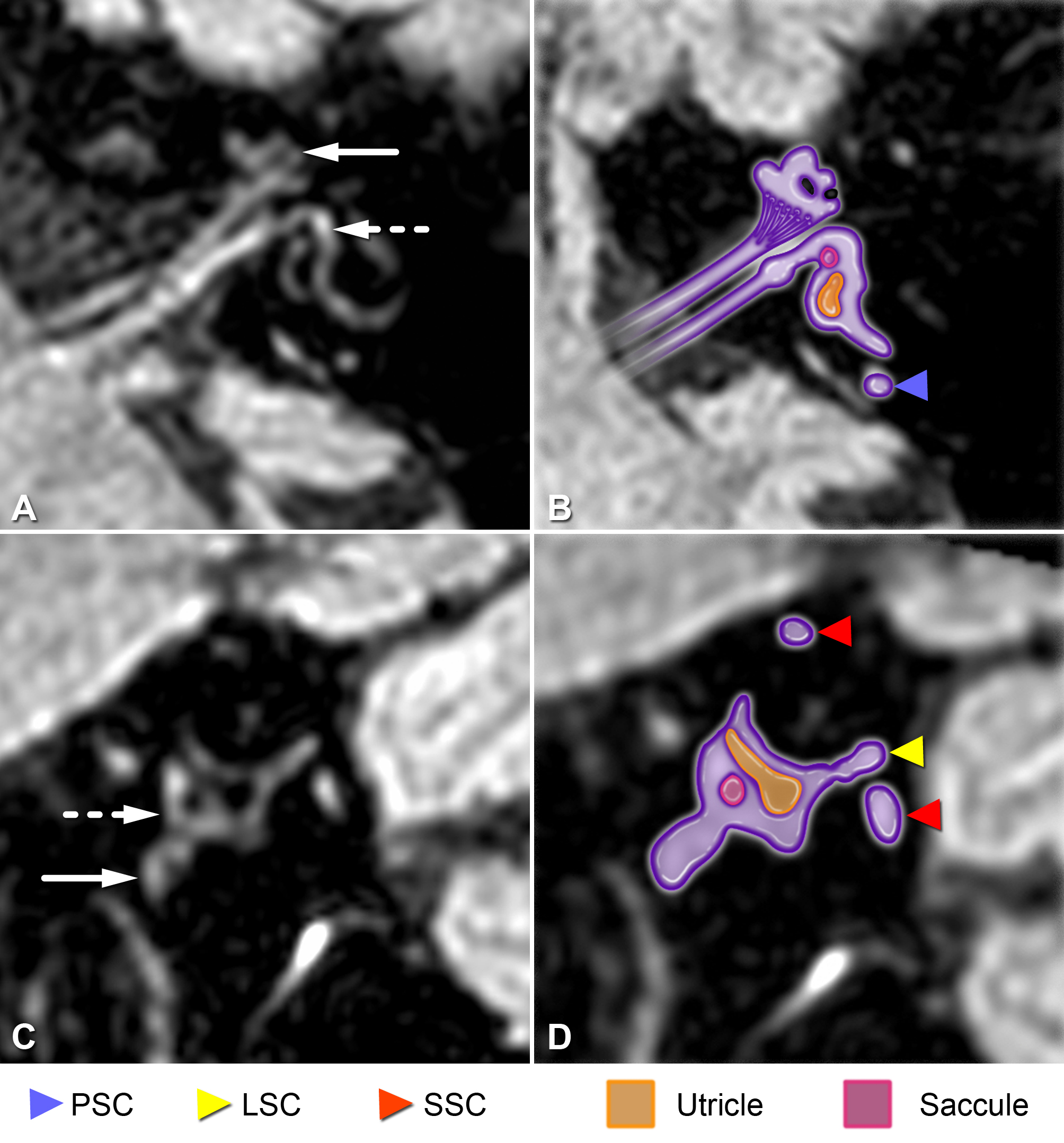
MRI Hydrops protocol in a healthy volunteer
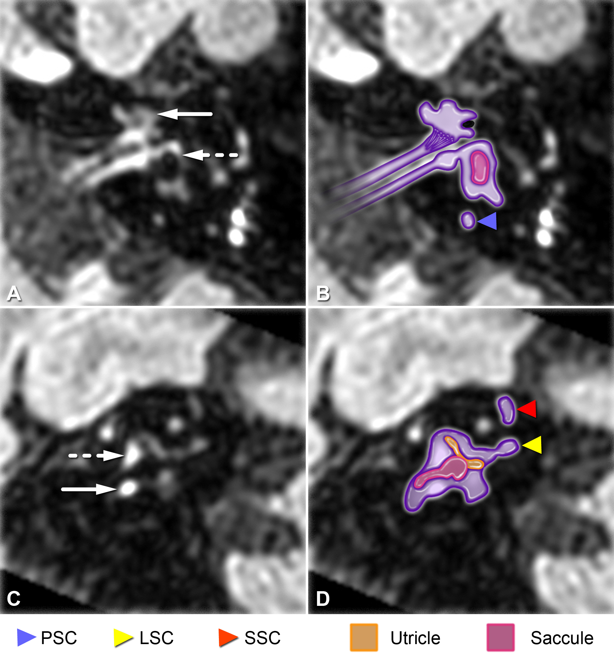
MRI Hydrops protocol in a patient with Meniere's disease
Publié le samedi 03 décembre 2016, 21:33 - modifié le 05/01/17 - Lien permanent